MetaMorph® Microscopy Automation & Image Analysis Software is the industry standard for automated microscope acquisition, device control, and image analysis, bringing microscopists greater understanding of cell morphology, function, and behavior for over 25 years. It is the ideal “glue” for easily integrating dissimilar fluorescent microscope hardware and peripherals into a single custom workstation, while providing all the tools needed to perform meaningful analysis of acquired images. The software offers many user-friendly application modules for biology-specific analysis such as cell signaling, cell counting, and protein expression.
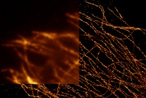
- Real-time super-resoution image processing supported by GPU hardware acceleration. Resolve sub-cellular objects as small as 20 nm spatially and 40 nm axially.
- Large set of supported hardware device drivers allows researchers to build custom imaging systems to meet their specific application need.
- Multi-dimensional acquisition (MDA) module allows users to easily perform complex acquisition sequences using a simple, guided user interface.
- Integrated morphometric analysis (IMA) measures and categorizes objects into discreet user-definable classes, based on any combination of morphometric parameters, such as shape, size, or optical density.
- Linked images, graphs, and tables allow users to easily view, classify, and correlate images with extracted data.
- Proprietary device and camera streaming accelerates image capture rate and simultaneously transfers images to memory during acquisition, capturing dynamic cellular events in applications such as live cell/kinetic imaging.
- Custom journals to further automate acquisition, processing, and analysis routines, created easily using a drag-and-drop interface on a graphical editor.
- 4D viewer with 3D measurements facilitates visualization of multi-dimensional data sets, stacks, and sequential images, such as multiple Z sections, wavelengths, time points, and positions. Multidimensional image data can be binarized into discrete objects for 3D isosurface viewing and rotation.
- Scan slide module automates acquiring multiple images larger than the field of view and then stitching them together. Ideal for large tissue samples, this ensures reproducibly while taking the guess work out of tiling experiments.
- Live replay captures real time events such as in FRAP, FRET and other laser-based studies, time lapse experiments, live cell imaging, and digital microscopy.
- Image enhancements such as standard kernel and morphometric filters, image arithmetic, and stack visualization, emphasize image characteristics that may not be discernable in the original image, making subsequent analysis and presentation more informative.