Real-Time Confocal or Widefield Fluorescence Image Deconvolution

The Microvolution software offers real-time deconvolution of images from widefield, confocal, two-photon, light sheet and HCA microscope systems. Microvolution can be integrated into the image acquisition workflow to allow us to provide seamless deconvolution during the capture of image sequences.

Image deconvolution removes the effects of signal noise and optical distortions in fluorescence imaging by using mathematical processing algorithms to restore contrast present in the original image. This processing intensive technique has been little used in recent years due to the length of time required to accurately generate the reconstructed image.

Developed by Stanford scientists, Microvolution harnesses the power of modern graphical processor systems to accelerate the analysis process and produce deconvolved images in a fraction of the time previously required. With analysis of datasets possible in seconds, it is now viable to integrate deconvolution into your image acquisition sequence.

Want to learn more...

  • Real time deconvolution of 2D and 3D fluorescence images
  • Almost instantaneous deconvolution (Up to 200x faster than competitors)
  • Accurate deconvolution
  • Dynamic analysis allows reduction in light levels to achieve same results with less photobleaching
  • Deconvolution enables cleaner, more accurate measurements – co-localisation, tracking, neurite lengths, intensities.
  • Blind & PSF based deconvolution options
  • Standalone plug-in module for image-J or Fiji
  • Blind deconvolution option for noisy applications
  • Multi-GPU option for very large image sets

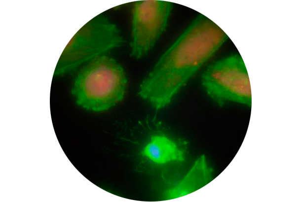
Before Deconvolution

Deconvolution of a 1024 x 1024 x 32 3-channel widefield fluorescence image, processed in 6 seconds total.

After Deconvolution​

Image courtesy of Dr. Dennis Hughes, Jared Mortus, Dr. Laurence Cooper, and Dr. George McNamara.