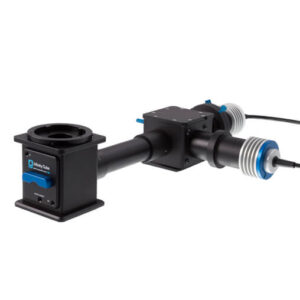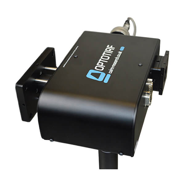

The OptoTIRF V2 is a compact and powerful, yet inexpensive, stepper-motor-controlled TIRF illuminator designed to fit onto any research-grade inverted microscope.
It allows a single or multimode laser spot to be focussed anywhere in the back-aperture of an objective lens with joystick or software control, and simple storage and recall of preset positions via digital or COM interface. This makes it suitable for acquisition protocols involving TIRF and / or oblique illumination at a range of penetration depths and wavelengths.
Although it lacks the fast scanning functionality of the Gataca iLAS (which we are also proud to distribute), the 360 degree stepper-motor-control does allow the user to illuminate from multiple points during an experiment, making it straightforward to tweak the illumination to the sample and minimise fringes or shading gradients. Field uniformity is further enhanced by a dither function that helps to avoid the artefacts associated with point TIRF. Flexibility is enhanced by a motorised bypass port, for widefield illumination using LEDs or a liquid light guide, or, for the addition of a second TIRF module for truly simultaneous dual-colour imaging.
Cairns’s design ethos is based on compatibility across a wide range of manufacturers. In the rare case where we do not have a compatible part, we have a custom design team available to provide a solution.
We provide a comprehensive 12-month warranty on all our products.
We aim for a delivery time of 2-4 weeks. However, the specific delivery time will be confirmed at the point of order.
We offer full training and support across our entire product range.
Yes, we have full custom design capabilities and a multidisciplinary team of scientists and engineers available to provide a solution that meets your needs.
Our CellCams are supported in Micro-Manager.