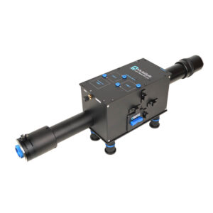
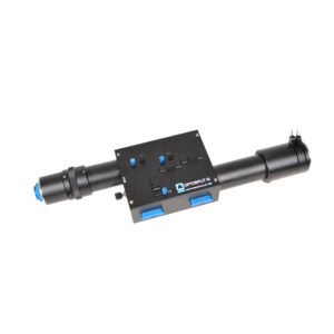
The Cairn OptoSplit II BP is our best ever dual channel simultaneous imaging device for use with a single camera. It builds on the success of the OptoSplit II image splitter, but adds a convenient single lever bypass mode making it more suitable for multi-user microscopes where simultaneous dual channels are only required for specific experiments alongside single wavelength recordings.
Whilst maintaining compatibility with the OptoSplit II, the BP version now supports our new flat-face filter cubes and has enhanced long-term stability, pixel registration and reproducibility. Featurewise, the rapid Bypass control is complimented by additional space for more auxiliary components. It has a slightly larger footprint than the OptoSplit II and consequently can use longer focal ratio lenses with even better off-axis performance.
Device drivers are included in most commercial imaging packages to assist registration and to allow real-time and off-line ratioing or fluorescence overlays. Alternatively the OptoSplit II BP can be used with simple image capture software and the processing carried out manually offline or using our own MicroManager and ImageJ drivers. The simple and accessible design makes the OptoSplit II BP an excellent platform for alternative applications, such as dual polarisation imaging.
Whilst optimised for coupling to a scientific microscope, the OptoSplit II BP image splitters can also be used with camera lenses or any other system of lenses which produce an image plane of suitable size and f/number (please ask for details).
Structuring Role of Tau-Tubulin Co-Condensates in Early Microtubule Organization. bioRxiv (Cold Spring Harbor Laboratory) — November 2024
Nanoscopic visualization of microgel-immobilized cytochrome P450 enzymes and their local activity — November 2024
Direct targeting of mitochondria by cisplatin leads to cytotoxicity in zebrafish lateral-line hair cells — October 2024
Whole-heart multiparametric optical imaging reveals sex-dependent heterogeneity in cAMP signaling and repolarization kinetics — January 2023
Dynamics of capillary blood flow responses to acute local changes in oxygen and carbon dioxide concentrations. Frontiers in Physiology — December 2022
DNA origami book biosensor for multiplex detection of cancer-associated nucleic acids — October 2022
Xanthene-stained nanoparticles for phosphorescence anisotropy measurements. Experimental Results — July 2021
Hot-Band Anti-Stokes Fluorescence Properties of Alexa Fluor 568. Journal of Fluorescence — May 2020
Microgel PAINT – nanoscopic polarity imaging of adaptive microgels without covalent labelling. Chemical Science — November 2019
Cairns’s design ethos is based on compatibility across a wide range of manufacturers. In the rare case where we do not have a compatible part, we have a custom design team available to provide a solution.
We provide a comprehensive 12-month warranty on all our products.
We aim for a delivery time of 2-4 weeks. However, the specific delivery time will be confirmed at the point of order.
We offer full training and support across our entire product range.
Yes, we have full custom design capabilities and a multidisciplinary team of scientists and engineers available to provide a solution that meets your needs.
Our CellCams are supported in Micro-Manager.