OptoSplit II

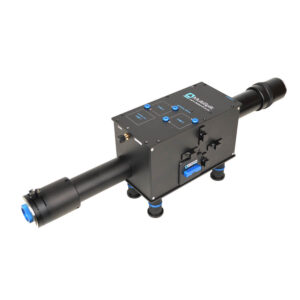
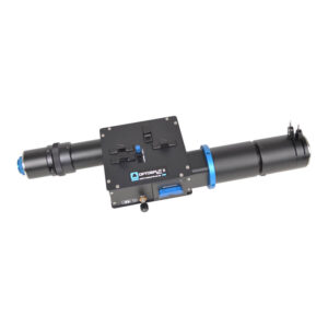
The Cairn OptoSplit II emission image splitter is a simple device enabling a single camera to record images simultaneously at two different optical wavelengths, polarisation states or other differentiated states.

Add to Quote
Share
Share
Share
Email

Traditionally, dual channel imaging is performed using an electronic filter changer or an additional camera and beamsplitter, neither of which is ideal for all applications. The switching speed of an electronic filter changer limits the temporal resolution, whereas a second camera adds cost and complexity. The OptoSplit uses a unique rotating mirror cradle, which gives adjustable spatial separation, to ensure excellent image registration and features a fully adjustable rectangular aperture to enable cropped sensor imaging modes and reduced scatter.

The OptoSplit uses our own proprietary lens design to support sensors up to 29.4mm diagonal. The instruments have a correspondingly larger aperture and improved off-axis correction to give enhanced performance with all sensors.

Device drivers are included in most commercial imaging packages to assist registration and to allow real-time and offline ratioing, or fluorescence overlays. Alternatively, the OptoSplit can be used with simple image capture software and the processing carried out manually offline, or using our own MicroManager and ImageJ drivers. The simple and accessible design makes the OptoSplit II an excellent platform for alternative applications, such as dual polarisation imaging.

Whilst optimised for coupling to any scientific microscope, the image splitters can also be used with camera lenses or any other system of lenses which produce an image plane of suitable size and f/number (please ask for details).

Application Usage

  • Förster Resonance Energy Transfer (FRET)
  • Ratiometric calcium, voltage & pH imaging
  • Simultaneous multi fluorescent probe imaging
  • Polarisation studies (anisotropy)
  • Simultaneous phase contrast / DIC and fluorescence
  • Simultaneous dual Z depth imaging
  • Total Internal Reflection Fluorescence (TIRF)
  • Spinning disk confocal
  • Single Plane Illumination Microscopy (SPIM)
  • 3D super resolution PALM/STORM (using cylindrical lenses)

Features and Benefits

  • Compact design with integral C-mount input and output ports as standard (F and T mount on request)
  • Support for sensors up to 29.4mm diagonal
  • Budget friendly alternative to two cameras
  • 425nm to 875nm AR coatings on all optical surfaces
  • 40mm diameter proprietary optics
  • Simple & precise controls for image registration
  • Interchangeable filter / dichroic holders
  • Emission filter dimensions – 25mm diameter
  • Recommended dichroic dimensions – 26x38x2mm (lambda/2 flatness)
  • Angled and flat auxiliary drop-in holders for 25mm ND filters and chromatic correction lenses (1 inch on request)
  • 1x 1.3x and 1.7x magnification available
  • Optional polarisation splitting kit including beamsplitter cube and rotatable polariser
  • Fixed or variable centre fully adjustable rectangular mask to delimit region of interest

Research Examples

Super-resolution localization microscopy visualizes ischemia-related cytoskeletal and vascular alterations at the nanoscale level in a mouse model of stroke. Neuroscience — December 2025

Single-Molecule FRET-Tracking of InlB-Activated MET Receptors in Living Cells — December 2025

Class A PBPs reinforce the septal cell wall following initial synthesis by SEDS–bPBP pairs during bacterial cytokinesis. bioRxiv (Cold Spring Harbor Laboratory) — September 2025

SlimVar for rapid in vivo single-molecule tracking of chromatin regulators in plants. Nature Communications — September 2025

Multi-parametric imaging of spatio-temporal cAMP signaling, transmembrane potential, and intracellular calcium in the Langendorff-perfused heart. bioRxiv (Cold Spring Harbor Laboratory) — April 2025

Effects of altered gravity on adrenergic-mediated cAMP signalling in intact cells. bioRxiv (Cold Spring Harbor Laboratory) — March 2025

Protocol for single-molecule labeling and tracking of bacterial cell division proteins. STAR Protocols — January 2024

A framework to validate fluorescently labeled DNA-binding proteins for single-molecule experiments. Cell Reports Methods — October 2023

Single-Molecule Localization Microscopy Using Time-Lapse Imaging of Single-Antibody Labeling. Current Protocols — October 2023

Single-Molecule Fluorescence Microscopy in Sensory Cilia of Living Caenorhabditis elegans. Methods in Molecular Biology — October 2023

Actin-generated force applied during endocytosis measured by Sla2-based FRET tension sensors. Developmental Cell — September 2021

Enzymatic studies on planar supported membranes using a widefield fluorescence LAURDAN Generalized Polarization imaging approach. Biochimica et Biophysica Acta (BBA) – Biomembranes — May 2017

Neuronal activity-mediated regulation of glutamate transporter GLT-1 surface diffusion in rat astrocytes in dissociated and slice cultures. Glia — July 2016

Molecular mechanisms underlying bile acid-stimulated glucagon-like peptide-1 secretion. British Journal of Pharmacology — February 2011

Retinal Oxygen Saturation in Patients with Systemic Hypoxemia. Investigative Ophthalmology & Visual Science — August 2011

Exploring membrane protein dynamics by multicolor single quantum dot imaging using wide field, TIRF, and hyperspectral microscopy. Proceedings of SPIE – February 2007

FAQ

Cairns’s design ethos is based on compatibility across a wide range of manufacturers. In the rare case where we do not have a compatible part, we have a custom design team available to provide a solution. 

We provide a comprehensive 12-month warranty on all our products.

We aim for a delivery time of 2-4 weeks. However, the specific delivery time will be confirmed at the point of order.

We offer full training and support across our entire product range. 

Yes, we have full custom design capabilities and a multidisciplinary team of scientists and engineers available to provide a solution that meets your needs. 

Our CellCams are supported in Micro-Manager. 

Customer Reviews

"The build quality of their equipment is excellent, and has proven both reliable and versatile in our lab’s day-to-day research; our Cairn equipment has provided years of dependable service."
Andrew James
“Over the past 20 years we have purchased a wide range of equipment from Cairn Research, ranging from several Optopatch amplifiers, OptoLED, Optosourse arc lamp and custom-made optical pathways for nanoscale measurements.”
Walter Marcotti
“What I love about this company is the quality and the reliability of every piece of equipment that I purchased. The instruments are made of high-grade materials (mostly aluminum for the optics components) and are intended for heavy everyday use in the laboratory. “
Andreas Werdich

Learn how we helped 100 top brands gain success

Cookie Overview

This website uses cookies so that we can provide you with the best user experience possible. Cookie information is stored in your browser and performs functions such as recognising you when you return to our website and helping our team to understand which sections of the website you find most interesting and useful.

You can adjust all of your cookie settings by navigating the tabs on the left-hand side.

For more detailed information on the cookies we use, please check our Cookie Policy.