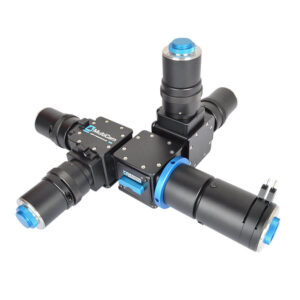
With the MultiSplit V2 the further possibility of simultaneous multidepth imaging is particularly attractive, as we can now do this at four depths rather than just two or three. In this application the splitting itself is on a neutral basis, but corrector lenses in the individual channels cause the focus to be at a different depth in the sample for each one, allowing the simultaneous acquisition of a four-channel “mini-z” stack.
Now we are four! Our OptoSplit and OptoSplit Bypass two-way image splitters and their TripleSplit (OptoSplit III) threeway cousin are now at last joined by our MultiSplit V2 fourway splitter. While a fourway design poses a greater technical challenge, which is why it took us a little longer to develop, it is potentially a very attractive product from a user point of view, as the four images are in a 2×2 square format rather than the linear rectangular one that the other splitters in our family generate. Furthermore, the continuing improvements in camera technology potentially allow the individual images to be acquired at megapixel resolution, which makes this form of image capture an increasingly viable alternative to the multiple camera approach, although of course we can also support that method if you prefer!
Just as with the other splitters in the Cairn family, the splitting can be performed on the basis of wavelength or polarisation, allowing applications where there is a requirement for simultaneous, or high speed, acquisition of multiple emission bands or polarisation states. The optical pathway is first split into two, and each of these pathways is then independently split a second time before refocussing to generate the four images. The simultaneous acquisition of these images offers a major benefit over manual or electronic filter changers, as there is no need to pause acquisition while the filter position is changed. This allows your camera to be operated in a fast streaming mode.
Whilst optimised for coupling to a scientific microscope, the MultiSplit V2 can also be used with camera lenses or any other system of lenses which produce an image plane of suitable size and f/number (again please ask for details).
Multiplexed imaging of G-proteins and ERK activity upon activation of CaSR. bioRxiv (Cold Spring Harbor Laboratory) — November 2025
Phosphoinositide lipids have bidirectional and spatially distinct roles in filopodial dynamics. bioRxiv (Cold Spring Harbor Laboratory) — October 2025
Nucleosome flipping drives kinetic proofreading and processivity by SWR1. Nature — December 2024
The chromatin regulator HMGA1a undergoes phase separation in the nucleus. ChemBioChem — November 2022
High-dimensional super-resolution imaging reveals heterogeneity and dynamics of subcellular lipid membranes. Nature Communications — November 2020
Cairns’s design ethos is based on compatibility across a wide range of manufacturers. In the rare case where we do not have a compatible part, we have a custom design team available to provide a solution.
We provide a comprehensive 12-month warranty on all our products.
We aim for a delivery time of 2-4 weeks. However, the specific delivery time will be confirmed at the point of order.
We offer full training and support across our entire product range.
Yes, we have full custom design capabilities and a multidisciplinary team of scientists and engineers available to provide a solution that meets your needs.
Our CellCams are supported in Micro-Manager.