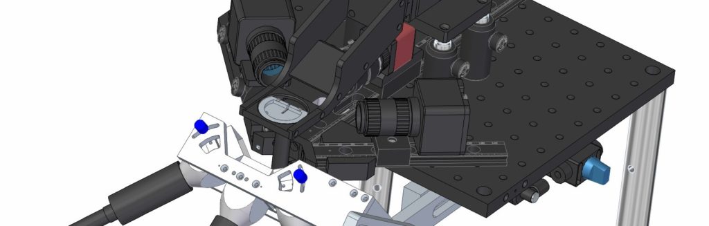
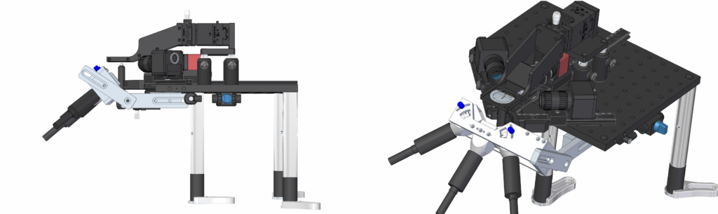
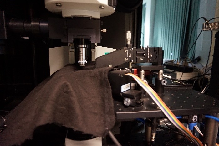
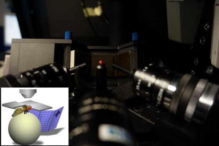
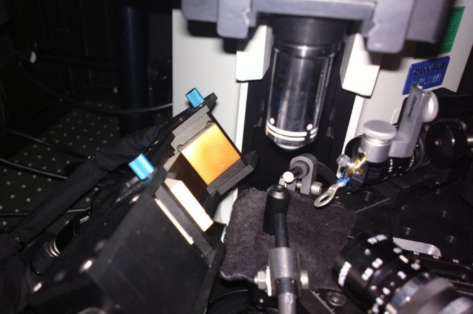
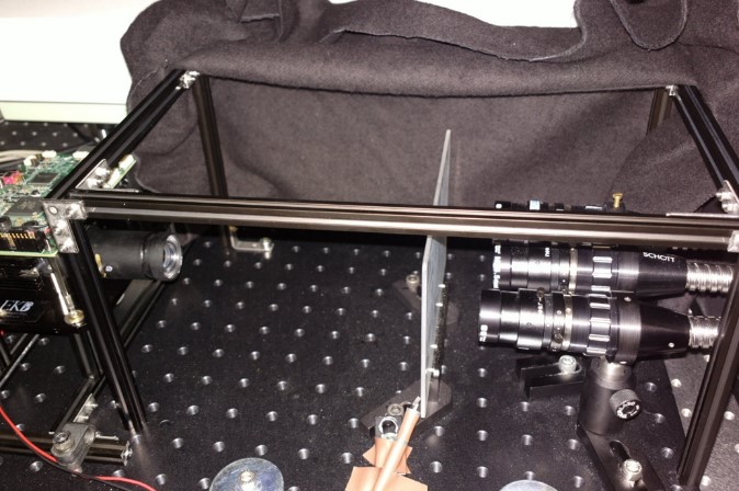
Being able to observe and link neuronal activity to behaviour in a moving animal is technically very challenging. Based upon the work of Dr Vivek Jayaraman (HHMI Janelia Research Campus, USA)[1], the Cairn Research Custom Microscopy Design team built a customised optomechanical assembly to fit the Juusola group’s multiphoton microscope. Here the head of the fruit fly is fixed, and its brain exposed, whilst the legs of the fly are able to move freely on a floating, rotating ball. This regime is referred to as ‘tethered walking.’ The ball (also called a ‘trackball’) is illuminated with infrared light and its motion, in response to the movements of the fly’s legs, is recorded by two cameras that form part of the optomechanical assembly. A third camera attached to the assembly permits the end user to precisely position the fly under the objective of the microscope for two photon calcium imaging, using GCaMP as the reporter. A novel, high-resolution projection system, designed and developed by the Juusola group was attached to the optomechanical assembly and could be precisely positioned to lie within the visual field of the fruit fly.
[1] Seilg, J. and Jayaraman, V. (2013), Feature detection and orientation tuning in the
Drosophila central complex,
Nature 503: 262-266