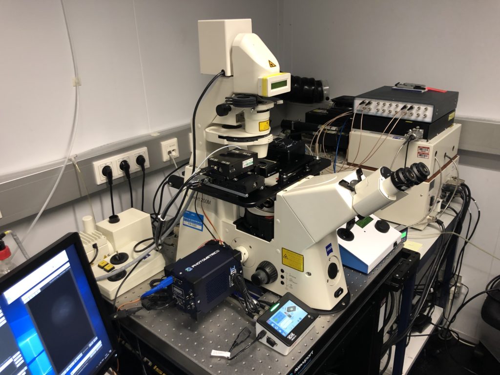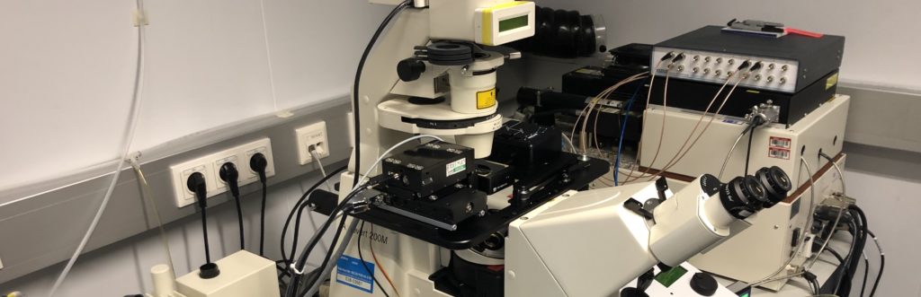
Imaging living specimens over an extended timeframe is challenging, owing to the increased risk of phototoxicity as well as the bleaching of fluorescent reporters. In conventional epifluorescence microscopy where the excitation light passes through the entire specimen, these risks are exacerbated; moreover, the visualisation of features of interest in the focal plane may be obscured by the excitation of fluorophores above and below it, known as ‘out of focus’ fluorescence.
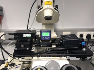
The group of Dr Kikuё Tachibana are interested in furthering understanding of how an oocyte transforms into a zygote following fertilisation. Insight into the mechanisms within the zygote that establish totipotency are also of great interest to the group, with chromatin architecture forming one area of focus. In order to gain such insights, the group relies on several advanced imaging techniques, including confocal microscopy. Oocytes and zygotes are difficult to image over an extended timeframe because they are extremely photosensitive; too much excitation light will arrest development. Therefore, a balance is essential. Images need to be acquired for a long enough time to capture sufficient signal and at a suitable frequency to ‘see’ and track the processes of interest, but not so frequently that the light dosage is such that it causes irreparable damage. For the reasons discussed above, this is particularly challenging using epifluorescence microscopy and the group therefore wanted to identify a new imaging modality better suited to their requirements.