 The various parts that need to be fitted to a microscope frame for phase contrast microscopy tend to be rather bulky. This classical arrangement for phase contrast imaging often restricts access to the sample from above on an inverted microscope frame, preventing the end user from applying certain experimental techniques. One such technique is Scanning Ion Conductance Microscopy (SICM) where an electrode is positioned immediately above the sample and can be used to map its surface profile without needing to be in direct physical contact with it.
Dr Gabriel Meloni (Warwick University) is interested in multimodal microscopy and wanted to find a way to combine SICM and phase contrast microscopy to monitor bacterial spore germination. It would not be possible to do this using a conventional phase contrast assembly on an inverted microscope frame because the component parts would hinder the precise positioning of the SICM electrode tip. Furthermore, the electrode assembly itself would block light from the transmitted light source reaching the sample, thereby degrading image quality.
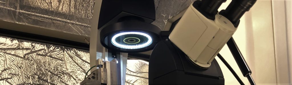
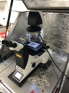
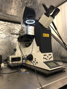 The Aura illuminator, which permits phase contrast imaging but can do so with fewer components versus the conventional configuration for phase contrast. For example, the condenser assembly is no longer necessary meaning that there is more free space above the sample to accommodate the SICM electrode body.
Within the Aura illuminator head there are several concentric LED rings, with each ring suited to either PhL, Ph1, or Ph2 phase contrast objectives. A custom adapter manufactured by Cairn Research enabled the Aura illuminator to be directly attached to the existing transmitted illumination pillar of the end user’s Leica DMI4000B microscope.