A very big thank you to Richard Wheeler from The Wheeler Lab, Nuffield Department of Medicine, University of Oxford for sharing his publication with us which uses our Multisplit for multifocal plane microscopy
Below is an animated gif of beating flagella recorded using our Multisplit with focussing lenses at the pupil planes to split the light to four quadrants of a single camera with focus offsets – Each plane is colour coded
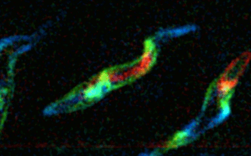
“The key advantage of using the Multisplit for multifocal plane microscopy was the ability to visualise the focal planes simultaneously. Other methods require some form of scanning, like scanning focal position by moving the sample/objective or scanning an illumination plane in selective plane illumination microscopy (SPIM). This will either introduce a shutter smear-like effect or severely restrict exposure time available. In contrast, the simultaneous capture of multifocal plane microscopy allowed the capture of small (single cell), faint (endogenously tagged proteins) and fast-moving (10s of Hz) subjects in 3D” Dr Richard Wheeler