EMBO Practical Course in Advanced Optical Microscopy – Plymouth, UK 2016
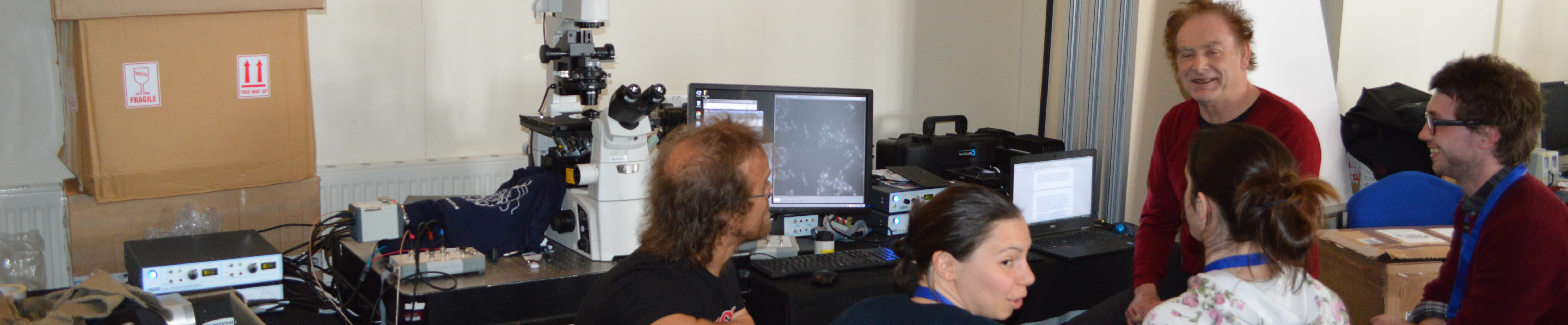
Twice a year Cairn travel to the MBA, Plymouth, UK to teach students microscopy related techniques. In September the course is called ‘Microelectrode Techniques for Cell Physiology‘ and in April it is the ‘EMBO Practical Course in Advanced Optical Microscopy‘. Martin, Danielle, Jez, Dan and Jim all travelled down to the 14th spring meeting with a complete imaging system fitted around a Nikon TiE illuminating fission yeast cells; The fluorescence illuminator was a dual channel OptoLED illuminating mNeongreen and mCherry, with a spinning filter wheel and image splitter in front of the camera on the emission side port.
We were demonstrating a solution to this problem – How can crosstalk be removed in fast simultaneous multichannel imaging?
When the green protein is excited with blue light, it will not only emit green fluorescence, but also some component of the same structure will bleed-through into the red channel. Now, imagine if the fluorescence of the green protein is much stronger than the red, then the crosstalk into the red channel from GFP could mask the much lower (desired) signal from the red fluorophore (RFP).
The only way to avoid this is to ensure that when the blue light is on, photons only reach the green detector area and that when the yellow light is on, photons only reach the red detector area, so there’s no cross-contamination between the two.
That’s very straight forward using conventional “slow” methods, because you would step in the appropriate illumination and detection filters between each measurement. The problem is if two or more emission filters are continuously in the light path, then you can’t avoid this contamination. Our fix is to synchronize a continuously spinning filter wheel with the camera so that different emission channels are in the light path sequentially within a single exposure.
The above text was taken from an article with our MD, Jeremy Graham
“The integrated OptoLED–OptoSplit–OptoSpin system gives the researcher confidence there is no cross-talk and the signal observed is solely generated by the expected fluorophore. Here the system is being used to examine the localisation of actin (mCherry labelled) and a myosin motor (mNeongreen labelled) in fission yeast” – Dr Dan Mulvihill – Universty of Kent at Canterbury