OptoScan Monochromator

• High throughput F/2 corrected optics
• Integral fast electronic shutter on all models
• New couplings using large silica-silica single core fibres

Product Description

Combined with our signal processing modules and optical hardware it forms the heart of a powerful standalone microphotometry system. Considerable effort has also been applied to making it the ideal illumination source for fluorescence imaging. At Cairn we see ourselves predominately as hardware designers and system integrators, so we have chosen not to develop our own imaging software and have instead sought to ensure compatibility with the excellent range of applications available on the market. Optoscan control is currently implemented in a wide range of commercial packages including MDS MetaFluor/Morph, RSI Neuroplex, Slidebook, Micromanager and Winfluor. We are able to provide turnkey solutions based on most of the applications below and have sufficient knowledge of all of them to offer comprehensive support. To summarise, if your application requires fast, flexible and automated illumination control then the Cairn Optoscan may well
be the instrument of choice.

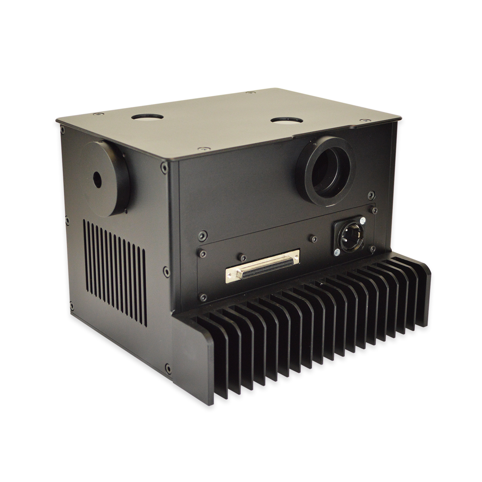
This instrument has been designed specifically with real-time biological fluorescence measurements in mind, but has evolved into a truly versatile laboratory tool. If your application requires fast, flexible and automated illumination control then the Cairn Optoscan may well be the instrument of choice.

Other commercial monochromators include the facility to change bandwidth between experiments using a manual control. This is sufficient for many purposes, but is not ideal for complex protocols especially those involving multiple fluorescence markers. Only by controlling bandwidth in real-time during experiments can each excitation wavelength be optimised independently. This means that where Stoke’s shifts are large or where fluorescence intensities are weak then a relatively large bandwidth can be selected to maximise signal-to-noise. Conversely if there is a small Stoke’s shift or if the fluorescence intensity is high then the slit width can be minimised to reduce bleed through and optimise dynamic range.


Download Data Sheet OptoScan Quick Start Guide (META) video support Get a Quote Back to Arc Light Sources

Additional information

Applications

• Fluorescence photometry
• Fluorescence imaging
• Optical scanning

Key Benefits

• Sub-millisecond control of centre wavelength with microsecond precision
• Sub-millisecond bandwidth control allows spectral and intensity optimisation at each wavelength
• Can be used with 75w lamps
• Modular design means that the light source can be removed and used independently as a white light source if required
• Light guide / fibre couplings to all popular microscopes
• External iris diaphragm for manual intensity control
• Built in fast electronic shuttering on all versions
• USB, Microprocessor, PCI, digital, and analog control option

Full Specification

• Optical configuration – enhanced Czerny-Turner configuration with fast F/2 light collection and off-axis parabolic mirrors to minimise
aberrations
• Diffraction grating – 1200 line ruled grating blazed for broad UV/visible range, 1800 and 2000 line holographic gratings for demanding
applications
• Wavelength resolution – 300-800nm specified with 0.5nm resolution @ ±1.5nm accuracy
• Bandwidth resolution – 0-30nm specified with 0.1nm resolution @ ±0.2nm accuracy
• Wavelength switching – 200nm transition <1.5ms, 50nm transitions in <1ms
• Bandwidth switching – Typical transition times of <1.5ms for both input and exit slits
• Digital shuttering (TTL) – Typically <2ms (depending on bandwidth)

Recent Publications

2023

2022

2021

2020

  • Chemogenetic analysis of how receptors for short chain fatty acids regulate the gut-brain axis – Read here

2019

  • Dynamic clamping human and rabbit atrial calcium current: narrowing ICaL window abolishes early afterdepolarizations – Read here
  • Microtubules Stabilization by Mutant Spastin Affects ER Morphology and Ca2+ Handling – Read here
  • Optogenetic stimulation of Gs-signaling in the heart with high spatio-temporal precision – Read here